FLOW VISUALIZATION IN PACKED BEDS
In our group, we have developed a technique of flow visualization using x-ray, exploiting the principle of x-ray absorption. A medical diagnostic x-ray unit is used to image the packed bed. The main aim of our flow visualization is to study the flow of non-wetting liquid in a packed bed.
Equipment Specifications
X-ray Unit: G.E. medical STENOSCOPE 9000
Liquid Medium: De-ionized water mixed with a dopant
Bed Type: Mono-sized particle packing
The flow of liquid is recorded on a computer/VCR and the recorded images are processed and stitched to give a complete picture of the bed under liquid flow condition. In the whole digitizing process, only one image processing operation has been used, thus reducing the errors substantially in quantification.
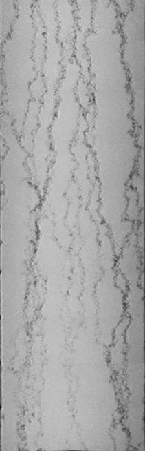
Results obtained from this technique have been verified using conventional methods and it has been shown that this method can give better results than the conventional methods. In Figure 1, one can clearly observe the liquid path (dark lines) through the bed, under the influence of gas, injected laterally. It can be seen that gas has pushed the liquid away.
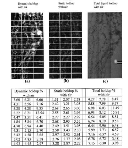
Various local holdups (static/dynamic/total) have been obtained in a liquid irrigated bed along with global holdups. Figure 2 shows the various local liquid holdups obtained in a packed bed in presence of gas flow, using this technique. Similarly local axial and transverse dispersion coefficients in a packed bed have been obtained.
This technique is being used to quantify the above parameters in presence of gas flow. A mathematical model is also underway to describe the dispersion phenomena in the packed bed based on above experimental findings.
For further details, queries, or publications related to this research, please contact us through our contact information on the website.